Fiberoptic & Rigid Bronchoscopy
Home / Dr. Kushal Chidgupkar
Interventional Pulmonology
- Fiberoptic & Rigid Bronchoscopy
- Endobronchial Ultrasound (EBUS) Guided FNAB & Staging
- Medical Thoracoscopy (Pleuroscopy)
- Endobronchial Stenting & Other Endobronchial Interventions
- Indwelling Pleural Catheter
- Tube Thoracostomy
- Intra-Pleural Fibrinolytic Therapy (IPFT)
- Chemical and Mechanical Pleurodesis
- Thoracentesis
- Trans Thoracic Biopsy (CT scan Guided Or USG Guided)
Fiberoptic & Rigid Bronchoscopy


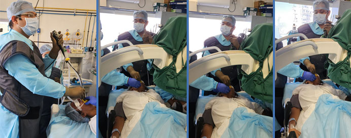
Bronchoscopy is a technique used to look at the air passages within the lungs with the help of a small camera that is located at the end of a flexible tube. This is connected to a video screen to view and record photos and videos. The tube also has an additional small channel to pass instruments within the airways for collecting lung tissue samples that can be used in disease diagnosis and also to carry out other interventional procedures.
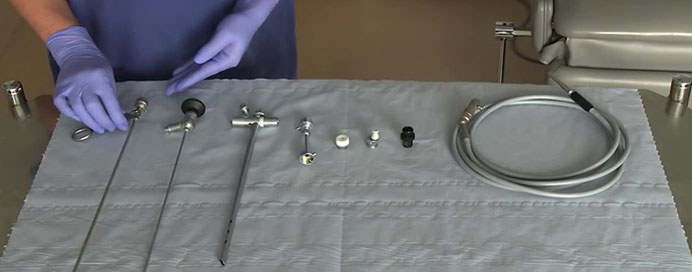
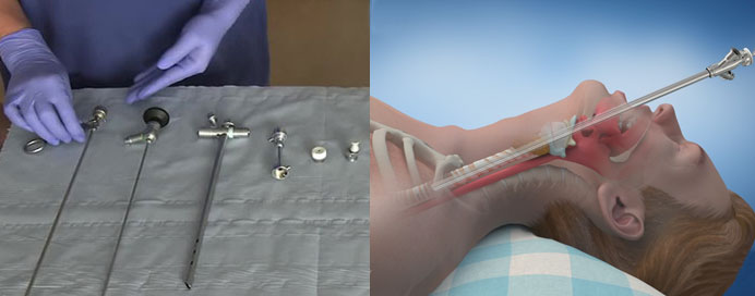
Bronchoscopy can be performed using a Flexible Bronchoscope or a Rigid Bronchoscope. More commonly used is the Flexible Bronchoscope.
Rigid Bronchoscope is used in situations where there is bleeding inside the lungs, in case of a large object stuck in the airways, or if some critical intervention is to be performed.
Bronchoscopy is done for diagnostic and therapeutic purposes. It is usually done to find the cause of the underlying lung disease. In case of persistent cough, fever or breathlessness associated with abnormal Chest X-ray or CT chest findings, one may have to undergo bronchoscopy to collect appropriate tissue samples to achieve the right diagnosis. This is particularly useful if we are not able to achieve the diagnosis by routine investigations. Also we can visualise the bronchial mucosa through the Bronchoscope. This helps us to have a better visual impression of the lungs and also to identify radiologically unseen abnormalities.

Broncho-Alveolar Lavage (BAL) is one of the most common techniques used to collect samples. This can be done by washing the affected areas of lung by instilling aliquots of sterile saline through the Bronchoscope and collecting secretions, mucous plugs through the suction channel of Bronchoscope. These collected samples are then sent for microbiological, molecular and cytopathological investigations so that we can identify the organism/s causing lung infection or the actual cause in non infective diseases.
Through the working channel of the bronchoscope we can pass forceps within the lungs and take biopsies. It can be either Endo-Bronchial Lung Biopsy (EBLB) which is taken through the larger airways in the central part of the lungs or Trans-Bronchial Lung Biopsy (TBLB) which is taken from the deeper peripheral areas of the lung. The biopsies can be done with the help of real time Fluoroscopy or Ultrasonography guidance (Radial Probe EBUS). To obtain tissue biopsies, Cup Biopsy Forceps are used commonly. Sometimes Alligator Forceps can also be used. We can also use Cryobiopsy Forceps to obtain bigger tissue chunks whenever required.
To control bleeding and resect Endo-bronchial growths, masses and fibrotic changes, Electrocautery, Argon Plasma Coagulation (APC) or lesser cautery can be used by passing specialised forceps through the working channel of bronchoscope.
In cases of Tracheal or Bronchial stenosis, or in cases where there is intrinsic or extrinsic compression of airways due to endo-bronchial masses, mediastinal masses, esophageal masses etc., occur significant narrowing of Airways or even complete obstruction may. This may lead to collapse of a part of the lung or even the complete lung, in case of which it can lead to severe breathlessness and desaturation, further leading to hemodynamic compromise. When a part of lung is collapsed, post- obstructive pneumonias can occur which can lead to complete destruction and bronchiectasis. If this airway narrowing is intrinsic then it can be treated using Electrocautery Knife, Argon Plasma Coagulation (APC), Laser Cautery Knife or many other forceps with the help of bronchoscope. Likewise, intrinsic or extrinsic airway narrowing can be managed with serial balloon dilations reduce space by passing specialised SRE Balloon Dilator through the working channel of Flexible Bronchoscope or Rigid Bronchoscope.
To keep the airways open, i.e to maintain the airway patency, thereby preventing further episodes of lung collapse, stenting of airways (Tracheal Stenting or Bronchial Stenting) is done. These stents can be either self expanding covered Flexometallic stents or Silicon stents. They can be introduced with the help of Flexible Bronchoscope or Rigid Bronchoscope depending on the type of stent used, or the location where stent it is to be placed.
Flexible Bronchoscope or Rigid Bronchoscope can be used for the removal of thick mucus plugs, broncholiths, foreign bodies which can lead to airway obstruction.
