Endo-Bronchial Ultrasound Bronchoscopy (EBUS)
Home / Dr. Kushal Chidgupkar
Interventional Pulmonology
- Fiberoptic & Rigid Bronchoscopy
- Endobronchial Ultrasound (EBUS) Guided FNAB & Staging
- Medical Thoracoscopy (Pleuroscopy)
- Endobronchial Stenting & Other Endobronchial Interventions
- Indwelling Pleural Catheter
- Tube Thoracostomy
- Intra-Pleural Fibrinolytic Therapy (IPFT)
- Chemical and Mechanical Pleurodesis
- Thoracentesis
- Trans Thoracic Biopsy (CT scan Guided Or USG Guided)
Endo-Bronchial Ultrasound Bronchoscopy (EBUS)

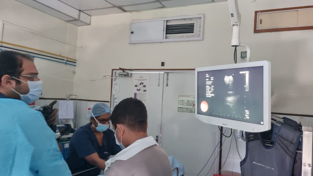
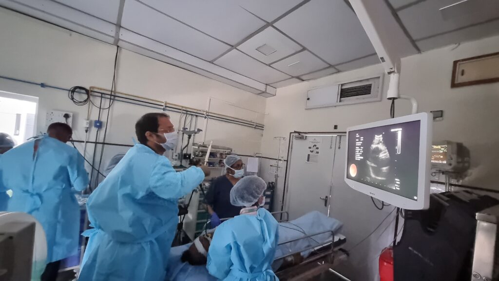
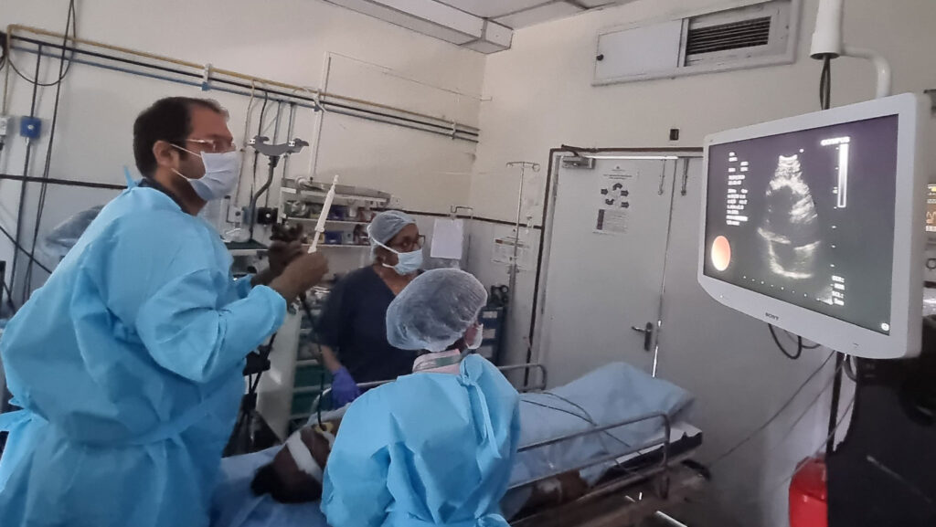
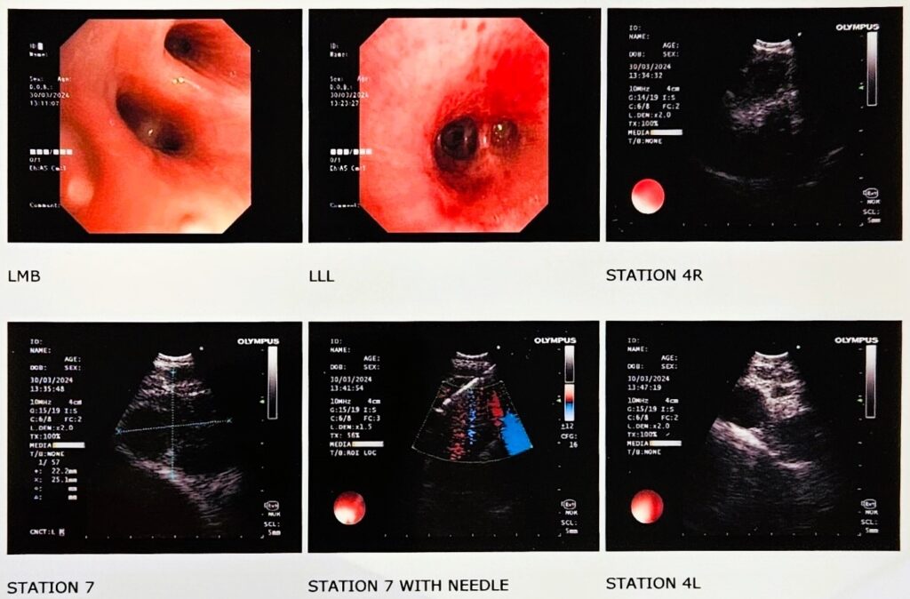
An Endo-Bronchial Ultrasound Bronchoscopy (EBUS) is a minimally invasive medical procedure done by Interventional Pulmonologist. By doing this procedure one visualises lymph nodes and mass lesions located adjacent to the airways. When EBUS technique was not available to visualise mediastinal lymph nodes, Mediastinoscopy was used. Mediastinoscopy is a more invasive procedure as compared to EBUS. In mediastinoscopy, a mediastinoscope is inserted through an incision at the top of the sternum.
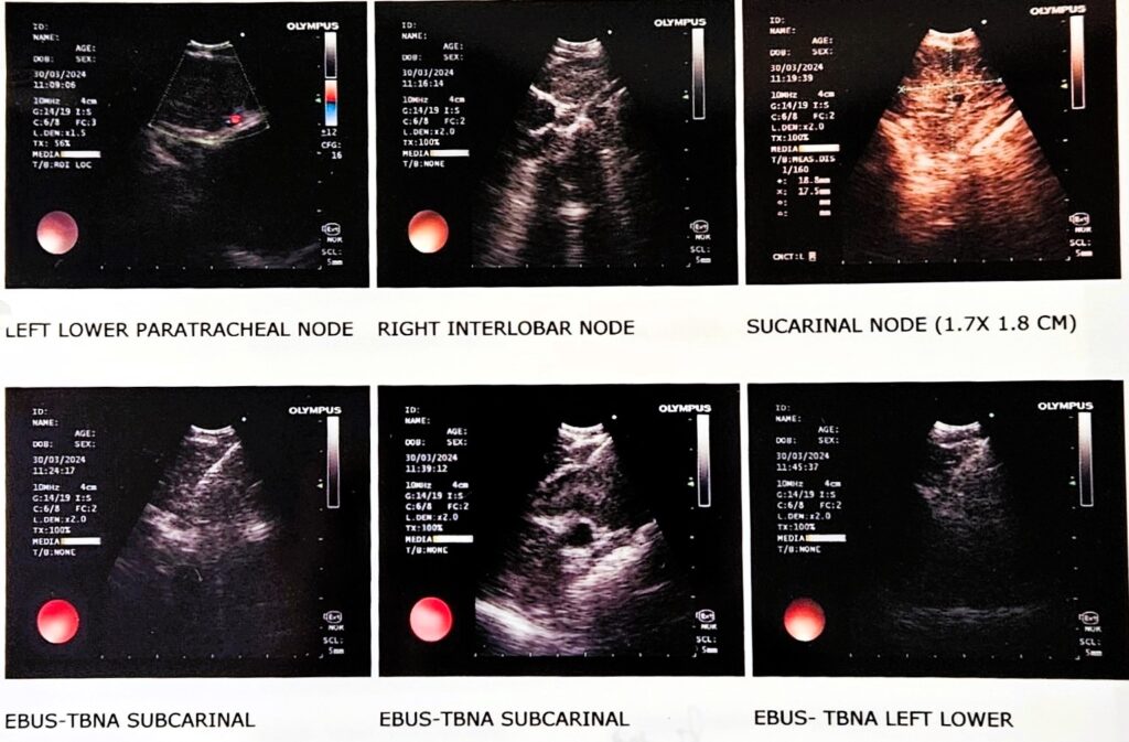
Endo-Bronchial Ultrasound is a bronchoscopic procedure. EBUS scope is made up of a specially designed Flexible Bronchoscope which has a camera and an USG probe at the tip. This scope also has a working channel to collect tissue biopsies.
Needle biopsy forceps are used to take tissue biopses i.e. Fine Needle Biopsy (FNB) and Aspirates (TBNA) from mediastinal lymph nodes and mass lesions adjacent to airways. These collected samples are sent for histopathological, cytopathological, microbiological and molecular investigations to find out whether patient is suffering from cancer, infection or inflammation.
Two different types of EBUS are available in the market- Linear (convex) EBUS and Radial EBUS. Linear (convex) EBUS is mostly used to visualise abnormalities in the central areas of lung and mediastinum. Radial EBUS is mostly used to visualise abnormalities in the distal areas of the lungs.
Radial EBUS gives a 360° view of the inner side of airways.
1- Benefits of EBUS
- Provides real-time imaging of the surface of the airways, blood vessels, lungs, and lymph nodes.
- EBUS provides better images which allow the Pulmonologist to easily visualise areas that are difficult to reach otherwise and also to access more and smaller lymph nodes for biopsy with the biopsy needle, as compared to the conventional mediatinoscopy.
- The accuracy and speed of the EBUS procedure lends itself to rapid onsite pathologic evaluation. Pathologists in the operating room can process and examine the biopsy samples as soon they are obtained and can request additional samples to be taken immediately if needed (ROSE: Rapid On SIte Evaluation).
- EBUS is done in a Bronchoscopy Suite and not in OT.
- It is performed under topical and local anaesthesia and moderate sedation.
- It is usually a day care procedure, thus patient can go home on same day.

