Medical Thoracoscopy (Pleuroscopy)
Home / Dr. Kushal Chidgupkar
Interventional Pulmonology
- Fiberoptic & Rigid Bronchoscopy
- Endobronchial Ultrasound (EBUS) Guided FNAB & Staging
- Medical Thoracoscopy (Pleuroscopy)
- Endobronchial Stenting & Other Endobronchial Interventions
- Indwelling Pleural Catheter
- Tube Thoracostomy
- Intra-Pleural Fibrinolytic Therapy (IPFT)
- Chemical and Mechanical Pleurodesis
- Thoracentesis
- Trans Thoracic Biopsy (CT scan Guided Or USG Guided)
Medical Thoracoscopy (Pleuroscopy)
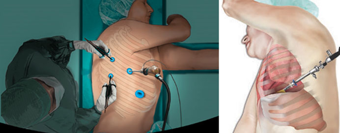
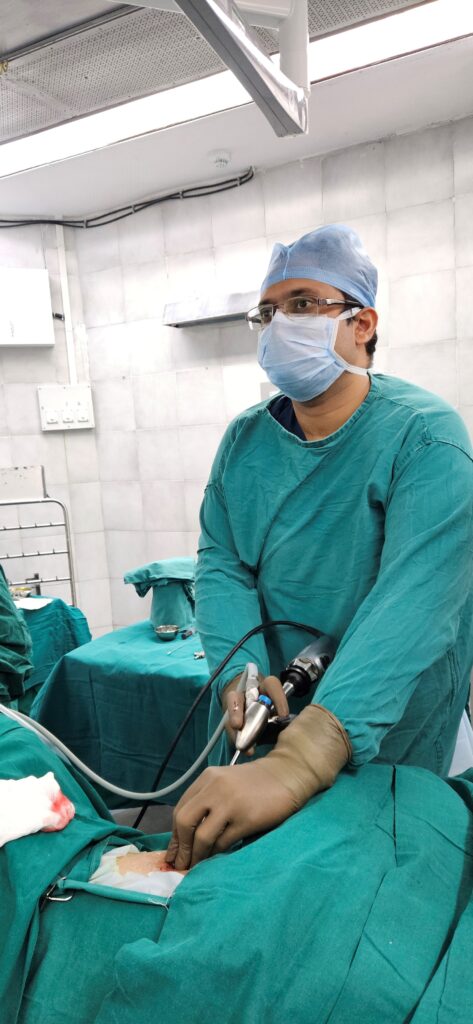
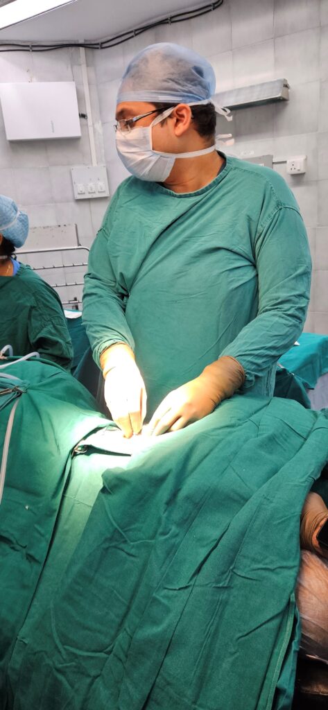
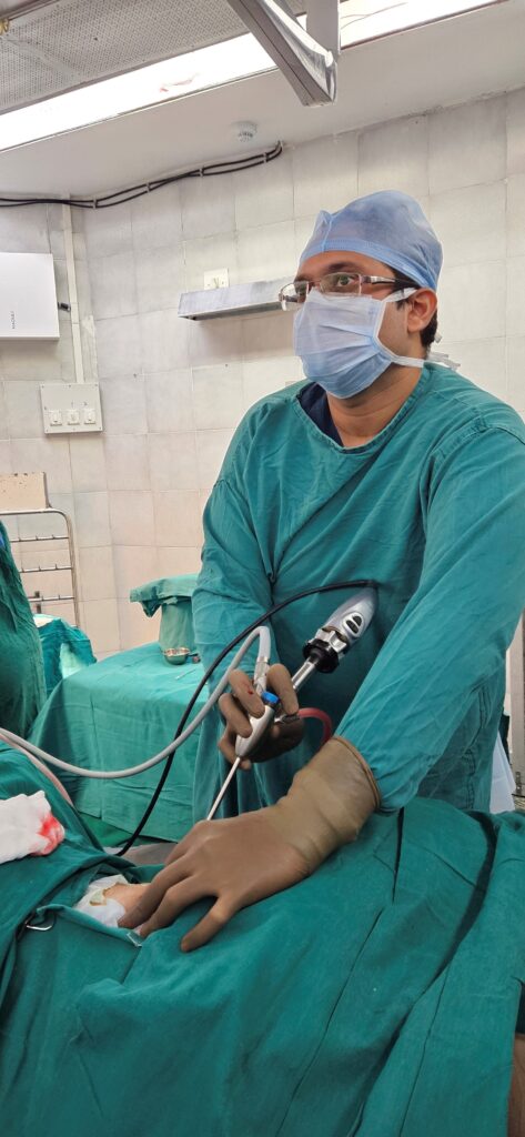
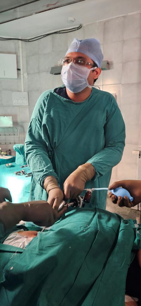
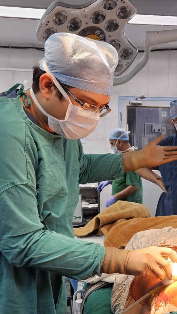
Medical Thoracoscopy is a minimal invasive procedure, performed to visualise the lung surface, pleura and the pleural space. It is performed by passing a medical Thoracoscope or Pleuroscope, through a small( about a centimeter) incision on the chest wall. Is a thin endoscope with a camera at it’s tip.
Medical Thoracoscopy is performed usually in the endoscopy room or minor operation theatre under topical anesthesia with mild to moderate sedation.
Medical Thoracoscopes are available in two types- Rigid Medical Thoracoscope and Flexirigid (Semi-Rigid) Medical Thoracoscope.
Rigid Medical Thoracoscope is the conventional one that is made up of metal. It can be inserted inside the pleural space through a single port opening for simple procedures and through multiple port openings for complex procedures. Now a days mini rigid Thoracoscopes are also available with better optics.
Flexirigid Medical Thoracoscope is a new advancement in medical thoracoscopy. It is made up of Fiber optic material same as Flexible Bronchoscope. It’s handle is similar to flexible Bronchoscope with a proximal rigid shaft and flexible distal tip which can be moved up and down for a better navigation.
Compared to surgical thoracoscopy, better termed as Video-Assisted Thoracic Surgery (VATS) which is performed in an operating room under general anaesthesia with elective intubation, medical thoracoscopy can be performed in an endoscopy suite under local anaesthesia or conscious sedation. Medical thoracoscopy is considerably less invasive and less expensive. As compared to VATS, the working spectrum and indications of medical thoracoscopy are limited to the pleural space only.

Indications of medical thoracoscopy
Thoracoscopic pleural biopsy is considered as the gold standard investigation for the diagnosis of the underlying cause of undiagnosed pleural effusions. Thoracoscopic Pleural Biopsy has a significantly high diagnostic yield i.e. >98%. In cases of isolated pleural based nodule we can target that nodule accurately under vision and achieve an appropriate diagnosis.
Performing adhesiolysis in loculated effusions: When pleural fluid remains in the pleural space for a long time, the fluid starts getting organized and loculated. Locules are formed due to septae forming pockets within the pleural space. These loculations cause the removal of fluid difficult. With the help of Thoracoscopy we can visualise the septations and also can pass different instruments through the Thoracoscope helping us to break such loculations easily.
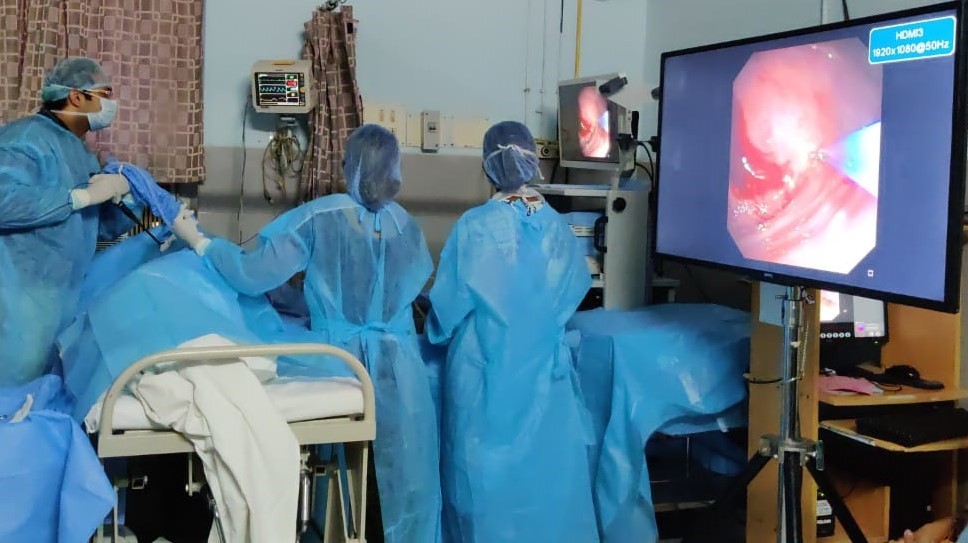
Pleurodesis: In case of recurring pleural effusions like malignant pleural effusions, thoracoscopy can be used to perform talc powder pleurodesis. During this procedure sterile talc is sprayed into the pleural space and this talc powder creates inflammation leading to the adherence of the parietal and visceral pleura thus preventing reaccumulation of fluid in Pleural space.
Staging of diffuse Malignant Mesothelioma and Lung cancers.

